Bird sperm is a tiny, elongated cell with a pointed head, a short midsection packed with mitochondria, and a long whip-like tail called a flagellum. You might be mixing this up with bird fleas, which are insect parasites, not microscopic sperm cells, and their appearance is very different what do bird fleas look like. The whole thing is somewhere between 30 and 300 micrometers long depending on the species, which means it is completely invisible to the naked eye. To actually see it, you need at least a basic compound microscope at 40x objective (400x total magnification), and even then you are looking at something smaller than a human hair is wide.
What Does Bird Sperm Look Like? Size, Shape, and Visibility

Marcus Chen
25 Jun 2026
Where bird sperm comes from
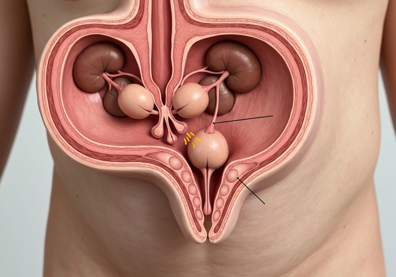
Male birds have a pair of testes tucked inside the body cavity near the kidneys, not hanging externally like in most mammals. That internal position is one of the more striking anatomical differences between birds and us. Each testis connects to a rudimentary epididymis and then to a highly coiled tube called the deferent duct, which runs alongside the ureter toward the cloaca, the shared opening at the base of the bird's body used for waste and reproduction.
During mating, the male everts his cloaca and transfers semen directly to the female's cloaca. There is no penetrative structure in most bird species. Once inside the female, sperm travel through the oviduct and get stored in specialized sperm storage tubules, where they can remain viable for days or even weeks depending on the species. So when you are thinking about what bird sperm looks like, you are thinking about cells produced in those internal testes, transported through that coiled duct system, and eventually released at the cloaca.
Why you can't see it without a microscope
Even though bird semen can be surprisingly concentrated, with turkey ejaculates containing over 10 billion sperm per milliliter, individual sperm cells are nowhere near visible without magnification. The cells are measured in micrometers. A micrometer is one-thousandth of a millimeter, so even a 300-micrometer sperm (the large end of the range) is only 0.3 mm from tip to tail. You would need exceptional eyesight and perfect lighting to notice anything at all, and what you would see would be a faint smear, not an individual cell.
Semen itself looks like a whitish or milky fluid, similar to what you might expect. what does a molting bird look like what you might expect. But the biological structure of any individual sperm cell, the head shape, the midpiece, the flagellum, those details only become visible under microscopy. This is true for most animals, birds included.
What bird sperm actually looks like under a microscope
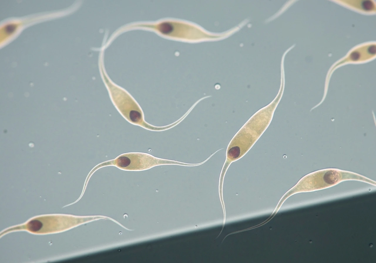
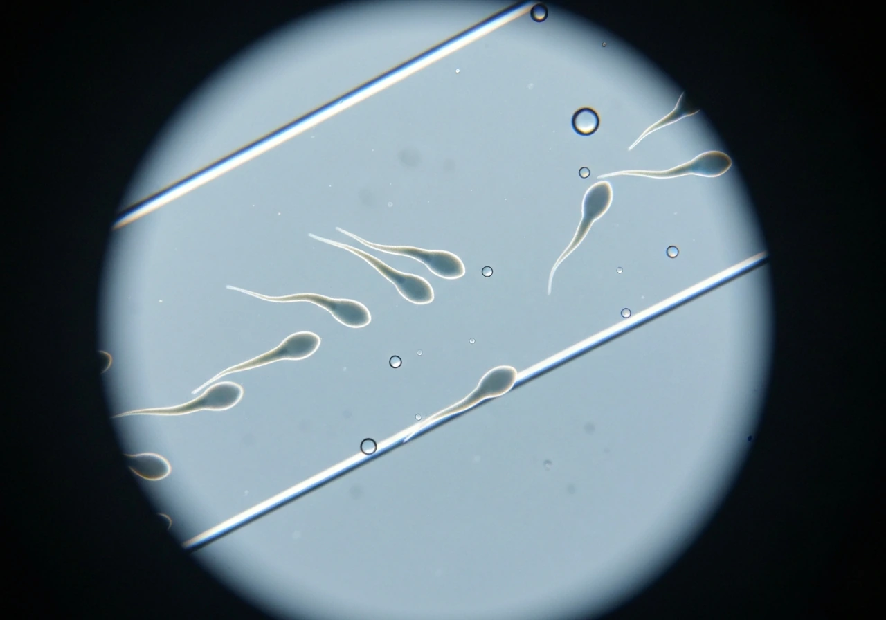
The general shape is consistent across most bird species: a narrow, elongated head at the front, a short midpiece in the middle, and a long flagellum (tail) that propels the cell forward. Researchers distinguish at least five structural regions in well-prepared samples: the acrosome (a cap at the very front of the head involved in fertilization), the nucleus (the bulk of the head, carrying the genetic material), the midpiece (where the mitochondria are packed to power movement), the principal piece (the main stretch of the flagellum), and a short endpiece at the very tip of the tail.
In motion on a fresh wet mount, bird sperm typically move in a fast, progressive, corkscrew-like pattern. The helical midpiece structure drives that spinning motion. This is especially noticeable in passerines (songbirds), where the mitochondria wrap around the midpiece in a helix. Motility is temperature-sensitive: studies in sparrows show that progressive motility drops and immotility increases when temperature rises above the optimal range, so a sample that has warmed up or dried slightly will look less active than a truly fresh one.
In terms of size, passerine sperm alone span roughly 40 to 290 micrometers in total length across species, a sevenfold range. For comparison, emu sperm (a large ratite bird) have a principal piece around 47 micrometers long, a midpiece around 3 micrometers, and a tiny endpiece under 4 micrometers. The head is narrow and pointed in most species rather than the oval paddle shape seen in human sperm. That elongated, almost needle-like head is one of the most visually distinctive features of avian sperm. If you are wondering about another bird species instead, like the yellowhammer, you can look up what a yellowhammer bird looks like in terms of color and pattern what does a yellowhammer bird look like.
How sample prep changes what you actually see
This is the part that trips people up. The same sperm can look noticeably different depending on whether the sample is fresh, fixed, or stained, and what kind of microscopy you are using.
Fresh wet mounts
A fresh sample placed on a slide, ideally in a shallow counting chamber like a Leja slide (about 20 micrometers deep), shows living sperm in motion. You get real-time motility but structural detail is limited because the cells are moving and unstained. Fast-moving sperm can actually be harder to examine morphologically because they are a blur before you can focus on them. You also have a short window before the sample dries or the motility drops.
Fixed and stained smears
Fixing the sample stops everything in place and lets you examine structure in detail. Common fixatives include formaldehyde, glutaraldehyde, and Hancock's solution, but these can cause some shrinkage, which means fixed sperm may measure slightly shorter than live ones. Different fixatives produce slightly different results, so if you are comparing measurements from two studies, the fixative used matters.
Staining makes structural regions much easier to distinguish. Eosin-nigrosin is a popular approach for live-dead assessment: live sperm appear white or pale against the dark nigrosin background because their intact membranes exclude the dye, while dead sperm absorb the eosin and stain pink or red. Fluorescent stains like SYBR-14 combined with propidium iodide take this further, labeling live sperm green and dead or membrane-compromised sperm red under fluorescence microscopy. Phase-contrast microscopy, which uses light interference to enhance contrast without staining, is another common approach and can resolve the acrosome, nucleus, midpiece, and flagellum regions clearly at 100x magnification.
| Prep method | What you see | What you lose or risk |
|---|---|---|
| Fresh wet mount | Live motility, real-time movement patterns | Short window before drying; structural detail limited on fast cells |
| Fixed smear (no stain) | Stable morphology for measurement | Shrinkage artifact; no live-dead info |
| Eosin-nigrosin stain | Live (pale) vs dead (pink/red) easily distinguished | Fine structural detail less visible |
| Phase-contrast microscopy | Head, midpiece, flagellum regions resolved without staining | Requires phase-contrast optics |
| Fluorescent SYBR-PI stain | Live (green) vs dead (red) with high contrast | Requires fluorescence microscope |
How to tell sperm apart from everything else in the sample
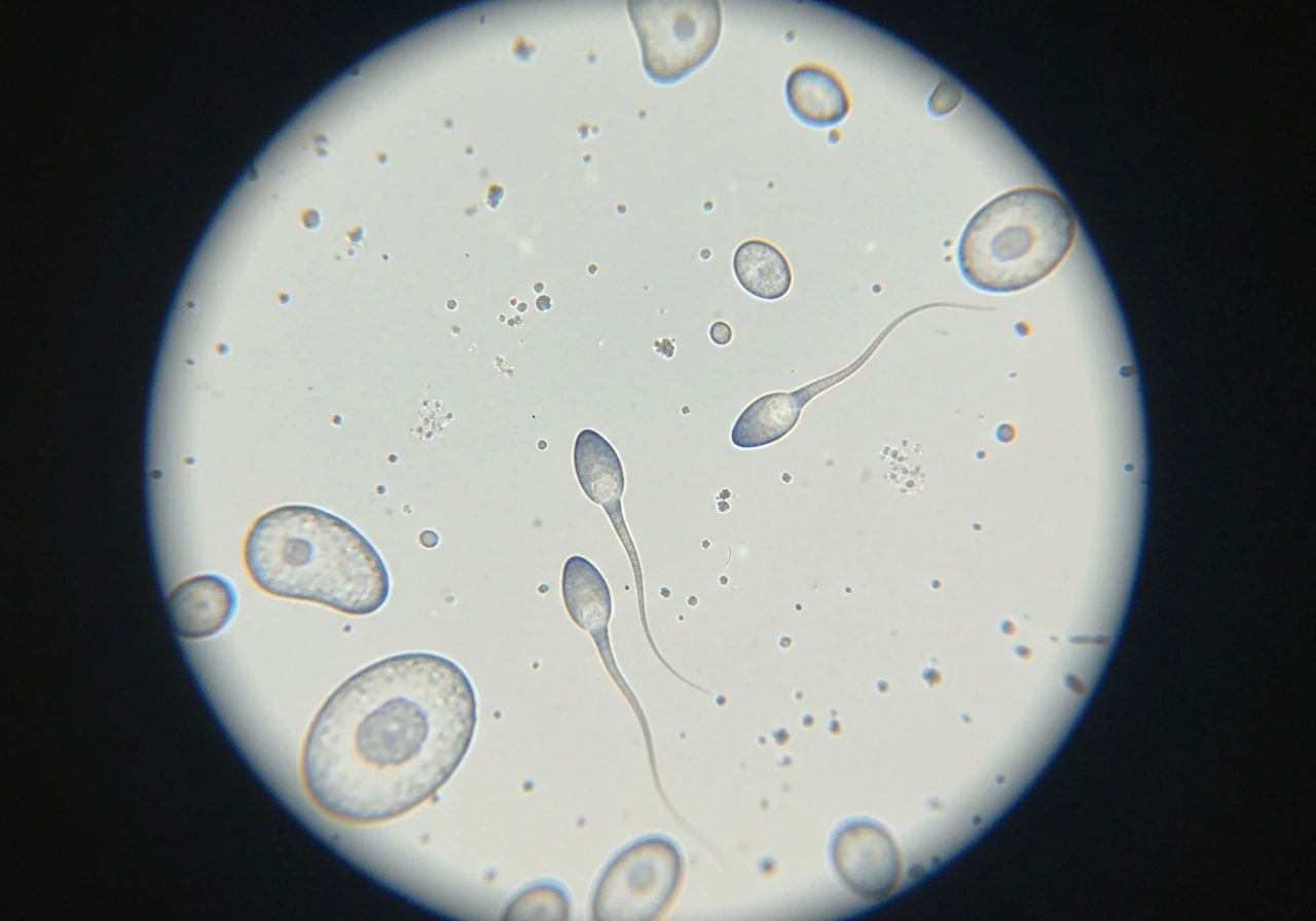
Semen is not a pure sperm solution. Seminal plasma (the fluid component) carries proteins, ions, and other cells, and under a microscope a raw sample has debris, epithelial cells, immature germ cells, and sometimes white blood cells. Bill law larvae insect bird pupa stage are separate topics from bird sperm, but the same careful microscopy-based approach applies when studying tiny life stages bill law larva insect bird pupa stage. Here is how to tell sperm from the noise.
- Shape: Mature sperm have a distinctly elongated, narrow head and a very long, thin tail. Most debris particles are irregular or roughly rounded with no organized flagellum.
- Movement: Sperm move with directed, progressive motion (often in a corkscrew pattern in birds). Debris drifts or vibrates randomly with Brownian motion. Nothing else in the sample swims purposefully.
- Tail swelling under hypotonic stress: In phase-contrast vitality testing, live sperm tails show a characteristic curling or swelling pattern in hypotonic solution. Debris does not respond this way.
- Staining response: Dead sperm and non-sperm cells stain differently with eosin-nigrosin than live mature sperm. Immature germ cells (spermatids) can look similar to sperm heads without the long flagellum and are a common misidentification risk with some staining protocols.
- Size consistency: Mature sperm from a given bird are relatively uniform in length compared to debris, which varies widely. If you see a population of similarly sized elongated cells with tails, those are almost certainly sperm.
The hardest calls are spermatids (immature sperm cells) and small debris particles. Spermatids have a rounded head and either no flagellum or a very short one, which distinguishes them from mature sperm. When using fluorescent stains, narrow structures like flagella can be hard to resolve and lead to undercounting or miscounting, so this is a known challenge even in professional lab settings.
Practical next steps if you want to observe bird sperm today
If you are genuinely trying to look at bird sperm, here is what you realistically need and what to expect at each level. Bird molting is the normal process by which birds shed old feathers and grow new ones, usually on a seasonal schedule.
- Get a compound microscope with at least a 40x objective (400x total magnification). A basic student-grade scope in the $150-$300 range can work. Phase-contrast optics give much better detail but cost more. You cannot use a simple pocket or dissecting microscope.
- Collect a fresh semen sample as quickly as possible and work with it immediately. Motility drops fast once the sample is outside the bird, especially if it cools or the seminal plasma breaks down. For pet birds like parrots, cloacal massage is the documented collection technique used in avian research.
- For a first look, place a small drop on a clean glass slide, add a coverslip, and observe at 100x then 400x. Look for elongated cells with long tails moving in a progressive corkscrew pattern.
- If you want structural detail, let a thin smear air-dry on a slide, then stain it. Eosin-nigrosin is accessible, does not require special equipment, and gives you a clear live-dead read alongside basic morphology.
- If the sample looks inactive, consider temperature: sperm motility in passerines is optimal around 38-40°C. A sample that has cooled to room temperature may show reduced or no progressive motility even if cells are intact.
- Compare what you see to published emu or chicken sperm images (freely available in journal articles) to calibrate your expectation for the narrow, elongated head and long flagellum typical of avian sperm.
How sperm varies by species, and what stays the same
The honest answer is that there is a lot of variation in sperm size across bird species, far more than you might expect. Total sperm length ranges from around 30 micrometers in some species to over 300 micrometers in others, with passerines showing roughly a sevenfold span on their own (about 40 to 290 micrometers). This variation does not track with body size in any simple way, which researchers find genuinely interesting. A tiny songbird can have proportionally very long sperm compared to a large raptor.
Despite that size variation, the fundamental architecture is conserved across virtually all bird species: a narrow elongated head with an acrosome at the tip, a mitochondria-packed midpiece, and a long flagellum. The midpiece organization, particularly how the mitochondria wrap around the cell in a helix, varies in detail between groups like songbirds and non-passerines, but the basic plan is the same. If you look at chicken sperm, emu sperm, and sparrow sperm side by side, you would immediately recognize them all as bird sperm because of that shared needle-like head and long whipping tail, even though the exact proportions differ.
Raptors, parrots, and ratites (like emus and ostriches) have all been studied in the context of conservation and captive breeding, so there is real published data on semen parameters for each group. Sperm concentration in raptors, for example, tends to be measured in the millions per milliliter range, much lower than domestic poultry. If you are working with a specific species and want to know what to expect, it is worth looking up species-specific semen parameter studies rather than relying on a single generic figure.
The broader takeaway: if you learn to recognize the core avian sperm structure (elongated head, midpiece, long flagellum, progressive corkscrew motility), you will be able to identify bird sperm across most species even without knowing the exact numbers. If you are also trying to identify a thrush in the field, it helps to learn the typical thrush look and key field marks for your region. The details vary, but the recognizable shape stays reliably consistent.
FAQ
Can I identify bird sperm from a picture, or do I need a microscope?
You usually cannot reliably identify bird sperm from typical photos because orientation and preparation strongly change how the head, midpiece, and tail appear. Even with magnification, images are often taken with a specific mounting method (wet mount, fixed smear, or stained prep), so the safest way to confirm is to know the sample type and look for the consistent avian features, a narrow pointed head, mitochondria-packed midpiece, and a long flagellum.
Why does bird sperm look different in “fresh,” “fixed,” and “stained” samples?
Fresh preparations emphasize living cell behavior, so sperm may look blurred during fast motion and structural detail is limited without dyes. Fixed samples preserve structure but can shrink or slightly distort measurements depending on the fixative. Stains add contrast that reveals regions like the acrosome and nucleus, but fluorescent labels can also make certain parts, especially thin flagella, harder to resolve and can affect counting.
What is the biggest reason people confuse bird sperm with something else?
The most common mix-ups come from looking at unclean semen or immature cells. Seminal plasma includes debris, epithelial cells, and sometimes white blood cells, which can mimic tiny shapes. Spermatids, which have a rounded head and little or no flagellum, are another frequent source of error because they can be mistaken for “small sperm” unless you use clear morphological criteria.
Is it possible to see bird sperm without a compound microscope?
In practice, no. Bird sperm lengths in the tens to a few hundred micrometers are far below what an unaided eye or simple magnifiers can resolve. Even simple “handheld” microscopes often cannot achieve the contrast and resolution needed to distinguish the narrow pointed head and midpiece organization, so you would likely see only an indistinct smear.
Does the motility pattern tell me anything specific about bird sperm health or freshness?
Yes, motility is a freshness proxy and it is sensitive to temperature and drying. If the sample is warmed or partially dried, you often see reduced progressive movement and more immotility. Also, sperm that are moving rapidly can be harder to assess for shape, so motility and morphology should be interpreted together rather than expecting perfect structural clarity in motion.
Can sperm storage in the female change what sperm looks like under a microscope?
It can change how they behave, even if the core architecture remains similar. Sperm stored in specialized tubules may show altered motility over time and may appear less active as viability declines, which can mislead you into thinking the morphology has changed. For accurate comparisons, you need to control storage duration and temperature conditions for the sample you are examining.
How can I distinguish mature sperm from spermatids or debris?
Mature sperm typically have an elongated, pointed head, a clear mitochondria-rich midpiece, and a long flagellum. Spermatids tend to have a rounded head and either no tail extension or a very short one. Debris can look irregular and lacks the conserved head-midpiece-tail layout, so focusing on the full three-part organization is more reliable than judging a single segment.
Why might fluorescent staining underestimate how many sperm you count?
Fluorescence can make thin, narrow structures like flagella difficult to resolve, especially if focus drifts slightly or if the imaging plane captures only part of the cell. That can lead to sperm being missed if your counting method relies on tail visibility. Using appropriate magnification, consistent imaging settings, and a counting rule that includes head-only recognition can reduce undercounting.
If I’m comparing measurements between two studies, what detail should I check first?
Check the sample preparation method and fixation protocol. Different fixatives and staining conditions can cause slight shrinkage and can also change how measurements are defined (for example, whether the tail is fully included or partially obscured). Without matching preparation and imaging criteria, apparent size differences may reflect methodology rather than true biological variation.