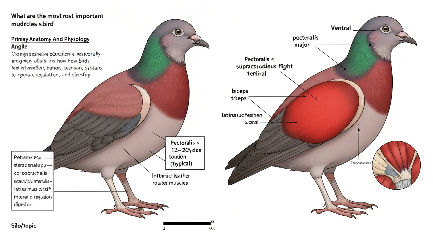
The two most important flight muscles in a bird are the pectoralis major and the supracoracoideus. The pectoralis is the massive chest muscle that powers the downstroke, and it is so dominant that it commonly makes up 12 to 20 percent of a flying bird's entire body mass. The supracoracoideus sits beneath it and drives the upstroke, routing its tendon through a bony pulley called the triosseal canal to pull the wing back up. Everything else, the shoulder stabilizers, the feather-tilting muscles, the elbow flexors, plays a supporting role to these two.
What Are the Most Important Flight Muscles in a Bird?

Marcus Chen
26 Jul 2026
Why bird flight muscles are worth understanding
I'll be honest: I used to think a bird just flapped its wings and flew. It seemed simple until I held a fresh-plucked chicken breast at a barbecue and realized I was holding the most powerful single muscle, relative to body size, of any vertebrate on the planet. That moment sent me down a research rabbit hole, and what I found was genuinely surprising. Bird flight muscles are not just large, they are precisely arranged, fiber-typed, and metabolically tuned in ways that explain everything from why a hummingbird can hover to why an albatross can glide for hours without burning much fuel at all.
If you are a student, a backyard birder, or just someone who watched a pigeon take off from a ledge and wondered how that works mechanically, this is the breakdown I wish I had found early on. No Latin walls, just the anatomy explained plainly, with numbers where they help.
The two primary flight muscles: pectoralis and supracoracoideus
Pectoralis major: the downstroke engine
The pectoralis major (often just called the pectoralis, or pect) is the primary power muscle for flight. It is the large, layered mass sitting on the bird's chest, and if you have ever noticed how deep a bird's breastbone protrudes, that bony ridge, called the keel or carina, is essentially a mounting surface for this muscle. The pectoralis originates broadly across the ventral (belly-facing) and lateral surfaces of the sternal keel and adjacent parts of the pectoral girdle. Its fibers converge into a tendon that inserts on the deltopectoral crest, a raised ridge on the underside of the humerus (the upper arm bone). When the pectoralis contracts, it pulls the humerus downward and forward, producing the powerful downstroke that generates most of the lift and thrust in flapping flight.
The keel exists specifically because of this muscle. Birds that don't fly, like ostriches and emus, have a flat sternum with no keel, because there is no massive pectoralis to anchor. The deeper the keel, the more surface area for muscle attachment, and generally the more powerful the flight. In strong fliers like pigeons and ducks, the pectoralis can represent anywhere from 12 to 20 percent of the bird's total body mass. Across all avian families, the range runs roughly 7 to 25 percent, with high-performance fliers and rapid take-off specialists at the upper end.
Supracoracoideus: the upstroke muscle with a clever pulley
Directly beneath the pectoralis, sandwiched between it and the sternum, lies the supracoracoideus. This muscle has a fascinating anatomy that most people, including me before I looked it up, would never guess. It also originates on the sternum and pectoral girdle, but its tendon does not take a direct path to the humerus. Instead, it travels upward through a small bony tunnel called the triosseal canal, formed by the meeting of three bones: the scapula (acromion end), the coracoid (acrocoracoid process), and the furcula (the wishbone, at its epicleidial process). The supracoracoideus tendon passes through the triosseal canal (a bony passage bounded by the scapula/acromion, coracoid/acrocoracoid process, and the furcula/epicleidial process) and acts as a pulley, this routing is conserved in crown birds and is critical for efficient humeral elevation and rotation The supracoracoideus tendon passes through the triosseal canal (a bony passage bounded by the scapula/acromion, coracoid/acrocoracoid process, and the furcula/epicleidial process) and acts as a pulley—this routing is conserved in crown birds and is critical for efficient humeral elevation and rotation.. The tendon loops through this canal like a rope over a pulley, then inserts on the dorsal (top) surface near the deltopectoral crest of the humerus. When the supracoracoideus contracts, it pulls the humerus upward and rotates it, elevating the wing for the upstroke.
This pulley arrangement is one of the more elegant mechanical solutions in vertebrate anatomy. A muscle sitting on the underside of the bird manages to lift the wing from above by redirecting its force through a bony canal. This triosseal canal anatomy is conserved across essentially all modern flying birds, which tells you how critical it is. The supracoracoideus is typically much smaller than the pectoralis, usually about one-fifth of it by mass, running roughly 1 to 3 percent of a typical passerine's or duck's body mass. But its role, and its proportional size, scales dramatically with flight style, as we will get to.
How these muscles actually power a wingbeat
Think of a single wingbeat cycle as two distinct phases: the downstroke and the upstroke. The pectoralis does most of the work. If you're interested in comparative biomechanics or human core training, see our brief explainer on do bird dogs work abs. In vivo studies using electromyography (EMG, which measures muscle electrical activity) and work-loop analysis (which measures actual mechanical power output) consistently show that the pectoralis performs the majority of positive mechanical work during the downstroke. Published power measurements from pigeon studies, for example, range from roughly 108 to 273 watts per kilogram of pectoralis muscle mass depending on flight speed, kinematics, and measurement method. That's a wide range, and researchers note it reflects how sensitive the output is to subtle differences in wing kinematics and how much elastic energy is being contributed by the supracoracoideus tendon.
That elastic energy point is worth dwelling on. EMG studies show the supracoracoideus is active during the upstroke, but in many species it also contributes during the transition from downstroke to upstroke by stretching and loading its tendon like a spring. When the upstroke begins, some of that stored elastic energy is released, reducing the amount of fresh metabolic work the pectoralis has to do on the next downstroke. It is a small but real energy-saving mechanism built into the muscle-tendon architecture of the shoulder. Scientists still work through exactly how much each species relies on this elastic storage versus fresh muscle contraction, so the precise balance varies and is an active area of research.
The supporting cast: shoulder and wing muscles
Once you move past the pectoralis and supracoracoideus, you are looking at a set of smaller muscles whose job is orientation, stabilization, and fine control rather than raw power. For a human-training analogy, see bird dog exercise which muscles to learn which stabilizer muscles are engaged in similar orientation and balance tasks. These are the muscles that determine exactly where the wing is pointing during each moment of the stroke, and they matter enormously for maneuverability even if they are not generating the bulk of the lift. For readers interested in core stability and balance exercises that support wing-like control in humans, see guidance on how many reps of bird dogs should i do to improve trunk strength and spinal alignment. For a human-exercise perspective on stabilization and control, see what muscles do bird dogs work, which explains how bird-dog exercises target the core, lower back, glutes, and shoulder stabilizers.
Key shoulder muscles
- Deltoideus (with scapularis and clavicularis subdivisions): These muscles run from the scapula and clavicular elements to the proximal humerus and are responsible for protracting (swinging forward) and elevating the humerus. They also contribute to humeral rotation and stabilize the shoulder joint during rapid direction changes.
- Coracobrachialis: Originates on the coracoid and inserts on the humerus, helping to adduct the wing (pull it toward the body) and retract the humerus. It works alongside the pectoralis during the downstroke.
- Scapulohumeralis: A smaller rotator muscle connecting the scapula to the humerus; it contributes to rotation and stabilization, much like the rotator cuff muscles in a human shoulder.
- Latissimus dorsi: A broad muscle on the dorsal (back) surface connecting the spine and synsacrum region to the humerus. It helps retract and depress the humerus and is particularly active during gliding and at the transition between stroke phases.
Elbow and forearm muscles
- Biceps brachii: Crosses the elbow joint and flexes the forearm (folds the wing). It also has a role in stabilizing the elbow during the stroke.
- Triceps (multiple heads): Extends the forearm and is critical for deploying the wing fully during the downstroke. In large soaring birds, the triceps helps lock the elbow in an extended position so the bird can hold its wings open without continuous muscular effort.
Intrinsic feather and follicular muscles
Attached to the base of each flight feather follicle are bundles of smooth muscle, tiny arrector, erector, and depressor groups that can shift individual feathers relative to one another during flight. Hieronymus et al. (2016) showed that avian intrinsic feather musculature and inter‑follicular smooth muscle bundles (arrector/erector and depressor groups, often annotated mnEPcm, mnDP, mnRP) connect feather follicles and coordinately alter feather orientation and wing morphing Flight feather attachment in rock pigeons (Columba livia): covert feathers and smooth muscle coordinate a morphing wing (Hieronymus et al. 2016). These are collectively called the intrinsic feather musculature, and they work together to morph the wing shape in real time, fanning feathers apart on the downstroke for a larger surface and rotating them to reduce resistance on the upstroke. Research on rock pigeons has shown that covert feathers (the smaller feathers covering the bases of the flight feathers) are also moved by smooth muscle and coordinate with primary feather movement to produce a continuously adjusting wing surface. The scale of control is remarkable: a single wing has dozens of individually controllable feathers.
Quick reference: major flight muscles at a glance
| Muscle | Location | Origin | Insertion | Primary Action |
|---|---|---|---|---|
| Pectoralis major | Ventral chest | Sternal keel and pectoral girdle (ventral/lateral) | Deltopectoral crest of humerus (ventral) | Downstroke (primary power) |
| Supracoracoideus | Beneath pectoralis, ventral chest | Sternum and pectoral girdle (dorsal/cranial) | Deltopectoral crest of humerus (dorsal) via triosseal canal | Upstroke elevation and humeral supination |
| Deltoideus (scapularis/clavicularis) | Shoulder region | Scapula and clavicular elements | Proximal humerus | Humeral protraction, elevation, rotation |
| Coracobrachialis | Shoulder/axilla | Coracoid | Humerus (ventral surface) | Humerus adduction and retraction |
| Scapulohumeralis | Dorsal shoulder | Scapula | Proximal humerus | Humeral rotation and joint stabilization |
| Latissimus dorsi | Dorsal trunk | Spine / synsacrum region | Humerus (dorsal surface) | Humerus retraction and depression; gliding control |
| Biceps brachii | Ventral upper wing | Coracoid and shoulder region | Radius (proximal) | Elbow flexion; wing folding |
| Triceps (heads) | Dorsal upper wing | Humerus and shoulder girdle | Olecranon of ulna | Elbow extension; wing deployment and locking |
| Intrinsic feather muscles | Wing surface (follicle bases) | Feather follicle connective tissue | Adjacent follicle tissue | Feather fanning, rotation, and wing morphing |
Suggested labeled images for this article
Good diagrams make this topic click immediately. If you are illustrating this article, here are the three views that would do the most work for a reader:
- Keel and ventral chest view: A frontal, slightly angled view of the sternum showing the keel depth, with the pectoralis and supracoracoideus layers peeled back sequentially to show their relative thickness and their shared origin surfaces on the keel. Labeling should highlight the triosseal canal position at the shoulder and the tendon path of the supracoracoideus routing upward and over to insert on the dorsal humerus.
- Wing cross-section showing muscle attachments: A cross-section through the proximal wing and shoulder joint, showing the deltopectoral crest of the humerus with both the pectoralis and supracoracoideus tendons inserting from opposite surfaces. This view makes it immediately clear why the supracoracoideus needs a pulley to reach the dorsal humerus from a ventral origin.
- Dorsal and ventral muscle map side by side: A simplified dorsal (back) and ventral (chest/belly) silhouette of a generic bird body, with color-coded muscle territories: pectoralis and supracoracoideus on the ventral view, latissimus dorsi and scapulohumeralis on the dorsal view, and deltoideus bridging both. A scale bar or percentage-of-body-mass label next to the pectoralis territory helps readers viscerally understand why this muscle dominates avian body composition.
Muscle mass, fiber types, and what they mean for flight performance
Size is not everything in muscle physiology, but it matters a lot. The pectoralis is the single largest muscle in most flying birds, routinely 12 to 20 percent of total body mass across typical volant (flying) families, with a range from around 7 percent in some slower fliers up to 25 percent in rapid take-off specialists. The supracoracoideus typically runs about 10 to 25 percent of the pectoralis mass in most flying birds, scaling nearly proportionally with body size across species. What's interesting from recent large-scale comparative data (over 600 species) is that while the pectoralis scales fairly predictably, the supracoracoideus shows much more variation between species, and that variation correlates strongly with flight style, especially diving and hovering.
At the fiber level, the primary flight muscles of flying birds are dominated by fast-twitch oxidative glycolytic (FOG) fibers. In plain terms, these are fibers that can contract rapidly (fast-twitch) but are also loaded with mitochondria and a rich blood supply so they can sustain aerobic energy production for extended periods. This is the opposite of what you find in, say, a sprinter's leg muscles (which are fast but fatigue quickly) or a marathon runner's (slow but sustainable). Birds essentially need both simultaneously: they have to beat their wings dozens or hundreds of times per minute, and they need to keep doing it for minutes or hours. FOG fibers are the evolutionary answer.
There are exceptions. Some deep muscle bellies or fibers in highly specialized soaring or diving birds shift toward slower, more fatigue-resistant profiles. And the metabolic implications are real: a bird with predominantly aerobic flight muscle can sustain flight for hours, migrating hundreds of kilometers on a single fuel load, while a bird with more glycolytic (less oxidative) fibers can generate explosive power for rapid take-off or short sprints but will fatigue faster. The architecture of each bird's pectoralis, both the mass fraction and the fiber type mix, is essentially a record of its evolutionary lifestyle.
How muscle anatomy differs across species
Hummingbirds: the hovering extreme
Hummingbirds are the outliers that every discussion of flight muscles eventually reaches. Their combined flight muscles (pectoralis plus supracoracoideus together) average around 26 percent of body mass across the family Trochilidae, with some studies reporting ranges up to 30 percent. That alone puts them far above typical birds. But the really striking difference is the supracoracoideus proportion: while in most flying birds the supracoracoideus is roughly one-fifth of the pectoralis by mass, in hummingbirds it can reach 40 to 55 percent of pectoralis mass. The reason is hovering. When a hummingbird hovers, the upstroke is not a passive recovery phase, it generates lift just like the downstroke. You need a very large, powerful upstroke muscle to do that, and the supracoracoideus is it.
At the cellular level, hummingbird flight muscles are extraordinary. In the rufous hummingbird (Selasphorus rufus), mitochondria occupy roughly 34.5 percent of muscle fiber volume, one of the highest values measured in any vertebrate. Their fibers are tiny in cross-section, packed with capillaries, and loaded with myoglobin (the oxygen-storing protein that gives red muscle its color). Oxidative enzyme activities (citrate synthase, cytochrome-c oxidase) are off the charts compared to most birds. All of this supports the highest mass-specific aerobic metabolic rates recorded among vertebrates, which is what you need when you're beating your wings 50 or more times per second and maintaining that for minutes at a time.
Raptors: power, precision, and burst capacity
Hawks, falcons, and eagles (collectively called raptors) have flight muscles tuned for different demands depending on their hunting strategy. Accipiters like Cooper's hawks, which pursue prey through dense vegetation in rapid bursts, have large pectoral muscles with a higher proportion of fast glycolytic fibers for explosive acceleration, at the cost of some endurance. Buteos like red-tailed hawks, which soar and glide for long periods before stooping on prey, show more oxidative fiber profiles in their pectoralis, supporting prolonged low-cost flight. Falcons like the peregrine have a profile somewhere between the two: enough glycolytic power for their famous high-speed stoops, but still capable of sustained powered flight. The secondary shoulder muscles in raptors are also notably developed because precise wing orientation during a stoop or a grab requires fine-tuned muscle control that raw pectoralis power alone cannot provide.
Albatrosses: built for efficiency, not power
Wandering albatrosses (Diomedea exulans) take the opposite approach. They spend months at sea, covering enormous distances over open ocean using dynamic soaring, a technique that extracts energy from wind gradients near the water surface. Their pectoralis, while still large, is proportionally less massive than in many flapping fliers, and their fiber profile is highly oxidative to support long, efficient flight. Crucially, the triceps and elbow-locking tendons in albatrosses are hypertrophied (enlarged and reinforced), allowing them to hold their wings fully extended with minimal muscular effort, essentially gliding for free energetically. The latissimus dorsi also plays a larger role in managing wing position during soaring. Albatrosses have been recorded flying for hours without a single flap, and the musculoskeletal anatomy reflects exactly that lifestyle: minimize flapping, maximize glide.
Wing-propelled divers and penguins: when upstroke must generate thrust
It's worth noting one more extreme. Penguins and wing-propelled diving birds like auks use their wings to swim, which means both the downstroke and upstroke generate thrust in a denser medium than air. In penguins, the supracoracoideus has been measured at roughly 35 to 50 percent of pectoralis mass, a much higher proportion than in typical flying birds, because underwater the upstroke cannot be a low-effort recovery phase. This is a striking parallel to hummingbirds and underscores the same principle: when the upstroke has to do real work, the supracoracoideus grows to match.
A few practical notes on measuring and monitoring flight muscles
Researchers typically measure pectoral muscle mass by dissection and wet weight, often expressed as a percentage of total body mass. In the field, ornithologists and veterinarians use a pectoral muscle score, a simple visual and tactile assessment of the muscle mass on either side of the keel felt from outside the feathers, to gauge a bird's body condition. A bird with prominent, rounded pectoral muscles scores high; a bird where the keel protrudes sharply with muscle wasting on either side scores low and may be malnourished, injured, or ill. This kind of hands-on assessment is a routine part of wildlife rehabilitation and banding work.
Clinically, the flight muscles are among the first to show the effects of illness or nutritional deficiency in birds. Muscle atrophy (shrinkage) from infection, poor diet, or disuse after injury reduces flight capacity directly and measurably. Conversely, birds preparing for long migration actively build pectoral muscle mass alongside fat stores in the weeks before departure, a process called premigratory hypertrophy. Both processes are visible in body condition data collected during banding and rehabilitation, making the pectoral muscle one of the most informative single indicators of a bird's overall health and flight readiness.
FAQ
What are the most important flight muscles in a bird?
The two primary flight muscles are the pectoralis (major) and the supracoracoideus. The pectoralis is the main downstroke (power) muscle; it originates broadly on the sternal keel and adjoining surfaces and inserts on the humerus (deltopectoral crest/ventral humeral surface). The supracoracoideus is the main upstroke elevator: it originates on the sternum/pectoral girdle, sends a long tendon through the triosseal canal and inserts on the dorsal humerus to lift and rotate the wing.
How do the pectoralis and supracoracoideus work mechanically during flight?
The pectoralis contracts to produce the powerful downstroke, generating most of the positive mechanical work. The supracoracoideus contracts to raise the wing during the upstroke; in many species it mainly tensions its tendon and can store/release elastic energy to assist the upstroke. The supracoracoideus tendon uses the triosseal canal as a pulley to change direction of force and rotate the humerus.
What is the triosseal canal and why is it important?
The triosseal canal is a bony passage formed by parts of the scapula, coracoid and furcula. The supracoracoideus tendon passes through it, acting like a pulley so the muscle—located ventrally—can elevate and rotate the humerus dorsally. This conserved pulley anatomy is crucial for efficient wing elevation in modern birds.
Which smaller muscles support wing movement and feather control?
Secondary shoulder/wing muscles include deltoideus (scapularis and clavicularis), coracobrachialis, scapulohumeralis, biceps brachii and triceps heads; these control humeral rotation, protraction/retraction and stabilize joints. Intrinsic feather muscles and inter‑follicular smooth muscle bundles adjust feather orientation and coordinate wing morphing (feather erection/depression) during strokes.
How do muscle masses and fiber types vary among birds and what are the implications?
The pectoralis is the largest single muscle and commonly represents ~12–20% of body mass in many volant birds (range ~7–25%). The supracoracoideus is often ~10–25% of pectoralis mass (≈1–3% body mass in many species) but shows greater interspecific variability. Flight muscles are dominated by fast‑twitch oxidative (FOG) fibres with high mitochondrial density and capillarity, supporting sustained aerobic power. Species with extreme demands (e.g., hummingbirds) have especially high mitochondrial volume and capillary supply, enabling very high mass‑specific aerobic rates and rapid wingbeat frequencies.
How do different flight styles affect muscle proportions?
Flight style strongly influences relative muscle size. Hovering birds (hummingbirds) have exceptionally large combined flight muscles (≈21–30% body mass) and a relatively larger supracoracoideus. Wing‑propelled divers and penguins have relatively enlarged supracoracoideus (sometimes 35–50% of pectoralis mass) because the upstroke must generate thrust. Long‑distance soarers (albatrosses) tend to have lower mass‑specific pectoralis but musculature optimized for sustained, low‑frequencystrokes; raptors have muscles configured for explosive power and maneuvering.
Next Articles

How Many Reps of Bird Dogs Should I Do? Sets and Progression
Learn bird dog rep targets, sets, hold times, and progression rules to match your fitness and keep perfect form.

What Muscles Do Bird Dogs Work? Targeted Movers and Form
Bird dog muscle guide: core bracing, glutes, back and shoulder stabilizers, plus pelvic control for anti-rotation
How Much Weight Can an Osprey Bird Carry: Numbers & Limits
How much weight can an osprey carry: typical prey 0.5–2 kg (≈30–100% body mass); anatomy, flight limits and evidence.